Insphero GravityTRAP Manuel utilisateur
Product Manual
GravityPLUS™
Hanging Drop System
www.insphero.com
ISP-06-001, ISP-06-010
www.insphero.com

GravityPLUS™ Hanging Drop System Manual
2
Contents
Introduction 3
GravityPLUS™ Hanging Drop System Components 5
GravityPLUS™ Plate 5
GravityTRAP™ Plate 7
Generating 3D microtissues 8
Additional materials required 8
Preparation 9
Hanging-drop formation 10
Transferring microtissues 12
Pre-wetting 12
Microtissue transfer 13
Medium exchange in the GravityTRAP™ Plate 15
Analysis and assays in the GravityTRAP™ Plate 16
Annex 1: Microscopy of microtissues 17
Annex 2: Preventing evaporation 19
Annex 3: Dosing and medium exchange in hanging drops 21
Annex 4: Microtissue harvest 23
Annex 5: Trouble-shooting guide 25
Annex 6: Step-by-step protocol for NIH/3T3 microtissues 27
Annex 7: GravityPLUSTM limited use label license 31
Version 6.0, July. 2015
451-0005-01-F

GravityPLUS™ Hanging Drop System Manual
www.insphero.com 3
Introduction
The GravityPLUS™ Hanging Drop System1
represents the most reliable, versatile and
complete platform for the generation, long-
term cultivation, observation and testing of
3D microtissue spheroids in 96 well format.
Each two-plate system consists of one
GravityPLUS™ Hanging Drop Plate (“GravityPLUS™
Plate”) and one GravityTRAP™ Plate.
InSphero uses this system for routine large-scale production of assay-ready
microtissues. For a list of available 3D microtissue models for ecacy and
toxicology studies, please refer to 3D InSight™ Microtissues on our website
at www.insphero.com.
Advantages of the GravityPLUS™ Hanging Drop System:
1. Robust hanging-drop spheroid formation using the GravityPLUS™ Plate
2. Straightforward spheroid transfer to the GravityTRAP™ Plate
3. Easy long-term growth, assay and observation in the GravityTRAP™ Plate
4. Protocols available for assays and analysis in the GravityTRAP™ Plate
1 The GravityPLUS™ Hanging Drop System, including GravityPLUS™ and GravityTRAP™ Plates and related technology, is protected by
several granted and pending patents world-wide.
GravityPLUS™ Hanging Drop System Manual
4
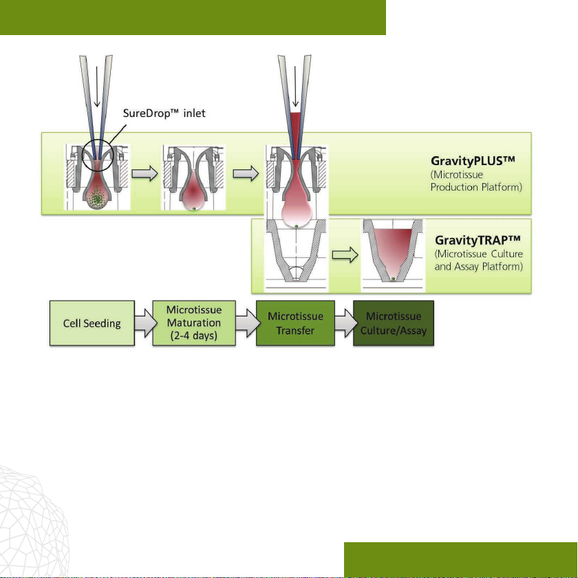
Figure 1: Microtissue formation in the GravityPLUS™ Plate and subsequent transfer to
the corresponding non-adhesively coated wells of the GravityTRAP™ Plate for further
cultivation and downstream applications.

GravityPLUS™ Hanging Drop System Manual
www.insphero.com 5
GravityPLUS™ Hanging Drop System Components
GravityPLUS™ Plate
The complete GravityPLUS™ Plate assembly consists of the following components:
1. Bottom plate (A) with reservoir (D)
2. GravityPLUS™ Plate (raster plate) with 12x8-well strips (B)
3. Lid (C)
4. SureDrop™ hanging drop 8-well strips (inserts, E)
5. Humidier pads (provided in bags with tweezers)
Figure 2: Components of
the GravityPLUS™ Plate

GravityPLUS™ Hanging Drop System Manual
6
Microtissue production with the GravityPLUS™ Plate is very simple. A cell
suspension is delivered from the top through the SureDrop™ inlet funnels of the
individual wells of the GravityPLUS™ Plate using a pipette or robotic liquid handler.
At the outlets under the plate, hanging drops will form and the cells will form
microtissues by gravity-enforced assembly within 2-4 days (Fig. 1).
After formation, microtissues are transferred into the GravityTRAP™ Plate. This
format facilitates long-term maintenance, optical visualization, compound dosing
and biochemical assays. If required, dosing and medium exchange can also be
performed directly in the GravityPLUS™ Plate - see Annex 3.
GravityTRAP™ Plate
The GravityTRAP™ Plate is a special non-adhesively coated 96-well microtiter plate.
It is designed to receive and accomodate microtissues for convenient long-term
cultivation and analysis. Microtissues are positioned in an observation chamber at
the bottom of each well, which prevents inadvertent aspiration and loss during
medium exchange (Fig. 3).
Biochemical assays as well as optical analytical methods such as brighteld and
uorescence microscopy can be performed. The GravityTRAP™ Plate ensures that
microtissues are centered in the optical viewing eld which enables automated
imaging processes (Fig. 4).

GravityPLUS™ Hanging Drop System Manual
www.insphero.com 7
Figure 3: GravityTRAP™ Plate – arrows indicating positioning pins for precise transfer of
microtissues from the GravityPLUS™ Plate.
Figure 4: HCT-116 colon carcinoma microtissue cultured
in the GravityTRAP™ Plate. Picture acquisition with a Zeiss
Axiovert25 microscope, 5× objective.

GravityPLUS™ Hanging Drop System Manual
8
Generating 3D microtissues
Generating 3D microtissues is a straightforward process that works with the vast
majority of cell types capable of forming tissues in vivo. In addition to the pro-
cess overview in this chapter, Annex 6 illustrates the formation of NIH/3T3 mouse
broblast microtissues in detail as an example for your own process.
Additional materials required
1. Mammalian cells, either cell lines or primary cells
2. 3D InSight™ Tumor Microtissue Media Kit (InSphero, cat. no. CS-17-001-01 -
includes 3D InSight™ Tumor Re-aggregation Medium (CS-07-111-02) and
3D InSight™ Tumor Maintenance Medium (CS-07-112-01)
3. Inverted microscope with a 5× objective or a 10× long distance objective with
a distance ring (see also Appendix 1)
4. Cell counter, e.g. Neubauer chamber
5. 8- or 12-channel pipette (e.g. Viao 10.0-300.0 μl, Integra Biosciences,
InSphero, cat. no. IS-001-01)
6. Medium reservoir for multichannel pipettes
7. For microscopic observation of the full GravityPLUS™ Plate, an additional
GravityPLUS™ bottom plate is recommended
8. Microplate centrifuge
9. Humidied CO2incubator

GravityPLUS™ Hanging Drop System Manual
www.insphero.com 9
Preparation
1. Wipe the GravityPLUS™ Plate bag with 70% EtOH before opening.
2. Carefully open the bag under sterile working conditions and take out the
GravityPLUS™ Plate assembly.
3. Prepare a reservoir (e.g. a 15 cm diameter petri dish) with 20 ml 0.5x PBS.
4. Open the bag containing humidier pads. Using the tweezers, remove one
humidier pad and place it in the dedicated reservoir containing the 0.5x PBS.
5. Wait until the humidier pad is completely soaked with PBS (approx. 5 min).
6. While pad is soaking, open the GravityPLUS™ Plate package and remove the
raster plate (Fig. 2-B).
7. Place the soaked humidier pad in the bottom plate (Fig. 2-A) of the
GravityPLUS™ Plate.
8. Trypsinize cells expanded in cell-culture asks according to your standard
protocol.
9. Count the cells.
Prepare a cell suspension. Recommended cell concentration: For long-term growth
proling start with low cell numbers (250–500 cells per drop). If non-proliferating
cells or rapid production of larger microtissues is required then start with 2500–
25,000 cells/40 µl.

GravityPLUS™ Hanging Drop System Manual
10
Hanging-drop formation
10. Gently deliver 40 µl of cell suspension into each well of the GravityPLUS™
Plate. It is easy to ensure tight contact between the pipette tip and the
well inlet by applying a slight pressure to form the SureDrop™ seal (Fig. 5).
Figure 5: Filling GravityPLUS™ wells. The pipette (8- or 12-channel) is positioned into the
inlet of the well in an upright or slightly tilted orientation. It is important that the pipette tips
make sucient contact with the well surface to assure complete liquid transfer and uniform
drop formation. The weight of the pipette alone is usually sucient to provide adequate
contact pressure.
IMPORTANT - For the homogeneity of forming microtissues it is essential
to assure a homogeneous distribution of the cells by gently pipetting up
and down prior to the seeding into the GravityPLUSTM Plate
Autres manuels pour GravityTRAP
1
Ce manuel convient aux modèles suivants
1
Table des matières
Autres manuels Insphero Équipement de laboratoire


Manuels Équipement de laboratoire populaires d'autres marques


Agilent Technologies
Agilent Technologies 5800 ICP-OES Manuel utilisateur

Endress+Hauser
Endress+Hauser Cleanfit CPA875 Manuel utilisateur

NI
NI PXI-5422 Manuel

Collomix
Collomix Aqix Manuel utilisateur

SPEX SamplePrep
SPEX SamplePrep 6875 Freezer/Mill Series Manuel utilisateur

Ocean Insight
Ocean Insight FLAME-NIR+ Manuel utilisateur


